Recent advances in fetal ultrasonographies have allowed these lesions to be diagnosed prenatally. These may be rounded flat or elliptical in shape.
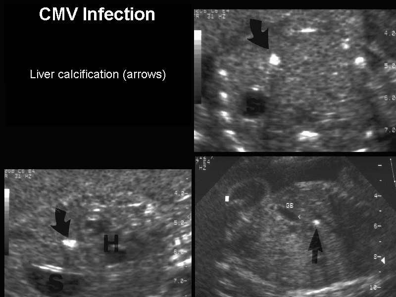
Hepatic Calcifications
To report our experience with the prenatal diagnosis of hepatic calcificationRoutine ultrasonography was done in 24600 consecutive pregnancies of 14.

. Hepatic calcifications in the fetus are hyperechogenic areas that are detected by ultrasonography imaging known as fetal liver calcifications FLCs. Fetuses with calcifications showed a significantly higher proportion of chromosomal abnormalities than controls. On my Level 2 ultrasound Im over 35 at 18 weeks 2 days the US tech saw a calcification on liver and then a bright bowel.
The incidence of chromosomal abnormalities and genetic syndroms is not increased. Objective The biological importance of calcifications occasionally noted in fetal tissues mainly liver at autopsy or ultrasound is largely unexplored. Parenchymal calcifications may be due to intrauterine infection.
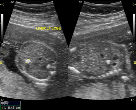
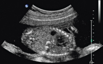
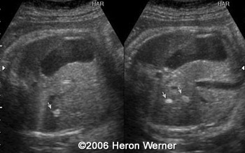
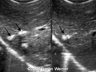
Organomegaly - abnormal enlargement of organs. The most frequent aberrations among cases included trisomy 21 33 trisomy 18 22 and monosomy X 18. A hepatic calcification is an area of abnormal brightness visualised within the fetal liver.
The purpose of this study was to provide information on the causes and postnatal outcomes of fetal liver calcifications that were detected by ultrasound imagingStudy Design. What causes a calcification in the. Signs of congenital CMV infection during pregnancy.
A detailed fetal ultrasound imaging for associated abnormalities maternal STORCH. Presence of solitary or multiple echogenic foci 1-2 mm in diameter within the substance of the liver or in the capsule. Fetal cardiac calcification is a rare ultrasound finding defined as diffuse hyperechogenicities affecting mainly the myocardium but can extend to involve the epicardium as well as the visceral pericardium.
All fetuses with isolated liver calcifications had a normal postnatal outcome 96 survival rate for fetuses with isolated. These lesions have been previously described as isolated findings or in association. Previous reports hint at an association to infection circulatory compromise malformations or chromosomal abnormalities.
How much did yours weigh around that time. I am currently 19weeks and 2 days ago had my anatomy scan where a 7mm calcification was detected in my babys liver. This fetus had cytomegalovirus and was the only fetus with increasing numbers of calcifications on follow-up scans.
The weight is 840grams and Im 26 weeks. This topic is answered by a medical expert. Everything was the same today.
Kate Smith answered this Calcification. Liver Calcifications are rare but are seen in about 1 in 1750 babies. Calcifications in the liver can be single or multiple and in most cases in which isolated hepatic calcific deposits are detected there is usually no underlying abnormality.
To identify factors associated with calcifications we have performed a case-control study on. I did the TORCH test and I am still waiting for the results. How does a Hepatic Liver Calcification happen.
If one or more of the following signs are identified via ultrasound an amniocentesis should be done to confirm a congenital CMV infection. Four of the 33 fetuses died one of which had liver calcifications as the only sonographic finding. The calcifications can be diffuse and involve the entire myocardium or patchy over large areas of the heart.
I have been worried and stressed since. The most frequent cause of focal calcified liver lesions is inflammation with granulomatous disease being the most common causeMost occurrences of granulomatous disease in the United States are attributed to histoplasmosis sarcoidosis and tuberculosis TB 24TB is one of the most prevalent causes of morbidity and death worldwide particularly in low- or middle-income. I am 22 weeks pregnant and my last ultrasound confirmed a calcification of the babys liver.
I have been referred to a fetal Medicine specialist with the possibility of having an amniocentesis. Typically seen as one or may echogenic foci within the liver. Calcifications were mainly located in the liver but also in heart bowel and other tissues.
Detailed sonographic studies amniocenteses and chromosomal bacteriologic virologic and serologic investigations were performed in each case with calcifications. Four of the 33 fetuses died one of which had liver calcifications as the only sonographic finding. The typical baby has only one calcification but some have more than one.
To report our experience with the prenatal diagnosis of hepatic calcification. The purpose of this study was to provide information on the causes and postnatal outcomes of fetal liver calcifications that were detected by ultrasound imaging. Calcium deposits on babys liver detected in ultrasound.
What causes a calcification in the babys liver and what should be my concern. Calcifications still there but apparently the growth is well and everything else is normal. Hepatomegaly abnormal enlargement of the liver.
In most cases the reason for the calcification is never found. By rpfeifer33954 1 post last post over a year ago. Im 26 weeks pregnant and recently had a level 2 ultrasound revealing a 4 cm spot white spot on my babys liver.
My daughter is 26 weeks pregnant and two calcium deposits have been detected on the babys liver. My doctor did not know what the concerns are. This fetus had cytomegalovirus and was the only fetus with increasing numbers of calcifications on follow-up scans.
Oct 19 2010 at 1116 AM. Routine ultrasonography was done in 24600 consecutive pregnancies of 14-26 weeks gestation. Dorsal echoes are usually absent.
The doctor is puzzled and doesnt know what this could be although he is thinking it probably isnt serious since its in the liver versus the heart brain bowels or kidneysThe ultrasound tech suggested it looked like a possible gall stone. Cases with fetal liver calcifications that were encountered between 1992 and 2001 were evaluated. Thirty-three fetuses had intrahepatic calcifications at 16-38 weeks gestation.
According to my doctor everything else looks alright. Outcome was determined after birth.
Fetal Intrahepatic Calcification Image Radiopaedia Org
Fetal Hepatic Calcification Radiology Key
Hepatic Calcification

Fetal Intrahepatic Calcification Radiology Reference Article Radiopaedia Org

Hepatic Calcification Radiologic Clinics

An Ultrasound Scan Of The Liver On Day 60 Showing An Enlarged Liver Download Scientific Diagram
Hepatic Calcification
Hepatic Calcification
0 comments
Post a Comment